- Research
- Open access
- Published:
Comparative evaluation and patient satisfaction with an electrical impedance-based device versus digital radiography in the estimation of remaining dentin thickness in carious posterior permanent teeth: (Diagnostic accuracy study)
BMC Oral Health volume 24, Article number: 429 (2024)
Abstract
Background
Accurate assessment of remaining dentin thickness (RDT) is paramount for restorative decisions and treatment planning of vital teeth to avoid any pulpal injury. This diagnostic accuracy study compared the validity and patient satisfaction of an electrical impedance based device Prepometer™ (Hager & Werken, Duisburg, Germany) versus intraoral digital radiography for the estimation of remaining dentin thickness in carious posterior permanent teeth.
Methods
Seventy patients aged 12–25 years with carious occlusal or proximal permanent vital posterior teeth were recruited. Tooth preparation was performed to receive an adhesive restoration. Pre- and post-excavation RDT were measured radiographically by two calibrated raters using the paralleling periapical technique. Prepometer™ measurements were performed by the operator. Patients rated their satisfaction level with each tool on a 4-point Likert scale and 100 mm visual analog scale (VAS). Inter and intragroup comparisons were analyzed using signed rank test, while agreement between devices and observations was tested using weight kappa (WK) coefficient.
Results
the intergroup comparisons showed that, before and after excavation, there was a significant difference between measurements made by both techniques (p < 0.001). After excavation, there was a weak agreement between measurements (WK = 0.2, p < 0.001), whereas before excavation, the agreement was not statistically significant (p = 0.407). Patients were significantly more satisfied with Prepometer™ based on scales and VAS (p < 0.001).
Conclusion
Prepometer™ could be a viable clinical tool for determining RDT with high patient satisfaction, while radiographs tended to overestimate RDT in relation to the Prepometer™.
Introduction
Successful restorative treatment of vital teeth depends on a holistic understanding of the tooth structure, mechanical and esthetic characteristics as well as function of the dentin-pulp complex which forms the necessary biological foundation for any restorative decision [1]. Since the best protective barrier for the pulp against trauma is the healthy dentin, it is very crucial during tooth preparation to consider the remaining dentin thickness (RDT). In the literature, an RDT of 2 mm is thought to be perfect for pulp protection [2,3,4]. Thin RDT less than 1 mm in deep preparations where more and wider dentinal tubules per mm2 are exposed, directly increases the risk for pulpal injury, affecting its repair response and subsequent restorative treatment options needed. Whilst in RDT of 0.5 mm, the dentin tubules are numerous, open, and wide enough as if there is a true pulpal exposure, [4]. Thus, it is very crucial to accurately assess the RDT especially in deep caries affected teeth to properly locate the caries excavation endpoint and decide the suitable restorative treatment thus preserving the tooth pulp vitality.
Clinical assessment of RDT is mainly dependent on integrating the operator’s knowledge of tooth anatomy and clinical experience, with radiographic findings interpretation [5]. However, this is very challenging owing to the subjective nature of these parameters. Although the radiographic assessment of RDT is the most available valid tool, it has many limitations such as the inconsistent estimation as Berbari et al., [4] found that it underestimated the real RDT by approximately 20% while on the contrary, Lancaster et al., [6] found that it overestimated the RDT. Besides its hazardous procedures to both the patient and dental personnel, overlapping of the anatomical structures and financial cost. Some other techniques have been investigated to assess RDT like laser fluorescence, pulse-echo, optical coherence tomography (OCT), and cone beam computed tomography (CBCT) however, they are not commonly used in dental practice for such a use [1, 7, 8]. Thus, the need for a real-time monitoring, non-invasive, user friendly and reliable tool is yet the most advocated.
One of the noninvasive clinical methods for RDT determination involves measuring electric resistance of the dentin by devices like Prepometer™ (Hager & Werken, Duisburg, Germany) and EndoEst 3D™ (Geosoft®, Russia), which is a multifunctional tool that combines a dentin meter to measure RDT, an apex locator and a pulp tester [9].
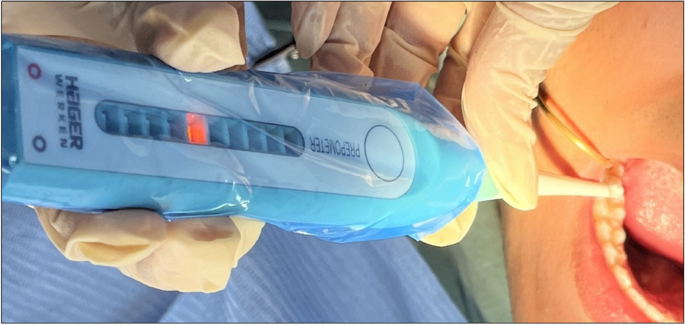
Prepometer™ is introduced to measure RDT only following tooth preparation. It depends on electrical impedance where an alternating 500-Hz electric current with an amplitude of 10A flows between a measuring sensory electrode placed on the prepared dentin and a reference electrode attached to the lip clip. The device also contains a third electrode for calibration. Ten LED lights of three colors like traffic light on the device measure, analyze, and show the resistance within ten seconds for each measurement. The light colors alter as the dentin electrical resistance declines, signaling an elevated danger of pulpal exposure [10].
Many invitro studies have used and evaluated the Prepometer™ [5, 9,10,11,12] nevertheless few clinical data are available; [8, 13] thus, this clinical study was introduced to help in proving the validity of this device clinically. Also due to the raised importance of patient-reported outcome measures (PROMs) and lack of sufficient knowledge in this area, patient satisfaction regarding the two devices was measured. The research question was that in patients with carious permanent posterior teeth, would the electrical impedance device be as valid as the digital radiography in clinical estimation of remaining dentin thickness? The proposed hypothesis is null.
Methods
Study settings
This clinical trial was conducted in Faculty of Dentistry- Cairo University, Egypt and was implemented following the ethical principles stated in the World Medical Association Declaration of Helsinki. The research protocol was reviewed and approved by the research ethics committee of the faculty with approval number (39–7-2022) on 26/7/2022. It was retrospectively registered on the clinicaltrials.gov website (https://clinicaltrials.gov/) with identification number (NCT06162182) on 8/12/2023. The trial has been reported following the STARD 2015 guidelines for Reporting Diagnostic Accuracy Studies.
Sample size calculation
A power analysis was designed by adopting an alpha (α) level of 0.05 (5%), a beta (β) level of 0.20 (20%), a within-subject correlation coefficient of (0.4), and a difference in proportions of (0.19) based on the results of a previous study [4] and on expert’s opinion; the predicted sample size (n) was found to be a total of (67) cases.
Clinical examination
Using a 0.5-mm ball-ended probe (CPITN Probe, Premium Instruments, USA) and a dental mirror, carious lesions were examined and scored according to the International Caries Detection and Assessment System “ICDAS”. Calibration of the examiners was executed using an online program on the International Caries Classification and Management System “ICCMS” website (https://www.iccms-web.com) to accurately define the eligible participants.
Eligibility criteria
Participants eligible for this study were those aged from 12 to 25 y, willing to join the study and with clinically detectable occlusal or proximal carious ( ICDAS score 3, 4) vital permanent posterior tooth with closed apex and healthy periodontal supporting. The minimum extension of the carious cavity should be at least 1 mm in width to accommodate with the probe of the Prepometer™ [14]. Excluded participants were those with poor oral hygiene, severe medical complications, showing signs and symptoms of irreversible or necrotic pulp pathology or with internal or external root resorption also when the affected tooth was with extended buccal or lingual caries, extending clinically very deep to the inner half of dentin thickness or previously restored. Also, patients suffering from any developmental or formative abnormalities e.g. molar incisor hypomineralization were excluded from the trial.
Patients were informed of the goals and procedures of the trial before consenting to participate and signing the informed consent form.
Operative procedures
All the operative procedures were performed under local anesthesia (articaine HCL 4% and epinephrine 1:100,000 (Artinibsa; Inibsa, Spain)) by one experienced operator. The tooth was prepared to receive an adhesive restoration. A tungsten carbide bur no. #245 (0.8 mm in diameter and 1.6 mm in length] (Komet, Germany) rotating in a high speed handpiece was used to remove the superficial and undermined enamel to gain access to the carious dentine and to remove caries from the walls for at least the 2 mm of the cavity boundaries to provide the peripheral seal necessary for the restoration success. The clinical determination of the caries removal endpoint was done based on the selective removal of caries consensus [15]. A sharp discoid excavator (#51&52 Dentsply Maillefer, Switzerland) was used to remove remaining carious dentine either to firm dentine “physically resistant to hand excavation and some pressure needs to be exerted through an instrument to lift it” in shallow and moderately deep cavities or to soft dentine “that deforms when an instrument is pressed into it and can be easily scooped up (e.g. with a spoon hand excavator) with little force being required” in deep and very deep cavities [15].
Measuring the remaining dentin thickness (RDT)
Index test: electrical impedance device, Prepometer™ (Hager & Werken, Duisburg, Germany)
The device was calibrated before any measurement by simultaneously touching the wet dentin surface with the calibration and sensor electrodes which is confirmed by the sequential flash of all the LEDs [10]. The reference electrode was placed on the buccal vestibule during the measurement. The sensor electrode was gently dragged across the cavity floor to measure the thickness of the remaining dentin at the deepest area, which was located by two trained raters and pointed by a periodontal probe [8, 14]. The electrical impedance value is displayed on the Prepometer™ by a scale represented by ten LEDs illuminated with different colors. They represent according to the manufacturer: green – a riskless preparation, yellow—further preparation is still possible, orange – limited range for a safe preparation and it should be stopped, finally red – imminent endangerment of pulp vitality [16]. Preoperative and post excavation measurements were taken Fig. 1.
Reference standard: intraoral digital radiographic examination
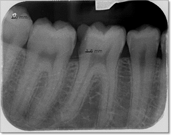
A preoperative digital periapical radiograph using the paralleling technique was taken of the selected tooth using an intraoral X-ray unit (Vario DG, Dentsply Sirona) with an exposure time of 0.88 s, at 70 kV, and a tube current of 3.5 mA using a photostimulable phosphor plate (PSP) film sensor of (31 × 41 mm) dimensions VistaScan® Imaging Plate PLUS, size 2 (Dürr Dental AG, Germany). Another post-excavation periapical radiograph using the same machine and settings was taken upon the completion of caries management. A digital reader preset (VistaScan Mini Plus, Dürr Dental, Germany) was used to process the radiographic images. The image analysis was executed using DBSWIN 5.4.0; a dental imaging processing software and visualized on a LED monitor (Lenovo D19-10 18.5 inch HD, China).
Two methods of image calibration were employed. First, the program was calibrated by equal matching the length of the imported radiographic photo to the actual length of the film sensor used for the study (41 mm). In addition, a reference guide was used which was the built-in (1 × 1 mm) triangle located at the corner of the used image plate. These calibration procedures allowed for millimeter-scale line measurements of the RDT. Two calibrated raters recorded all the measurements to confirm the reproducibility of the readings and to minimize the possibility of errors. They were blinded to the readings of the electrical impedance device, which were recorded by the operator. The radiographic image and raters’ calibration processes were done on ten initial cases with the help of a dental radiologist. The measurement was executed by drawing a straight line from the deepest point of the carious tissue floor to the highest point of the pulp before and after ending of the tooth cavity preparation [17] Fig. 2.
Patient-reported outcome measures (PROMs): patients satisfaction
Patients were evaluated regarding their satisfaction with the handling and conveniency of each device using a 4-point Likert scale: (a) “yes, very satisfied”, (b) “yes, mostly satisfied”, (c) “less satisfied”, (d) “not at all satisfied”. They were also asked to mark the Visual Analog Scale (VAS), which is a 100 mm straight horizontal line with the left end representing “not at all satisfied” while the right end indicating “very satisfied”. The satisfaction value was calculated by measuring the distance from the left end of the scale to the mark in millimeters and expressing it as a percentage (10 mm equals 10%, 20 mm equals 20%, etc.) [18].
Data treatment and statistical analysis
In order to obtain common units for comparison and based upon a previous study [19], electrical impedance device “Prepometer™” green LEDs were representing 2.1 to 3.0 mm radiographic RDT, yellow LEDs to 1.5 to 2.1 mm, orange LEDs to 0.9 to 1.5 mm while red LEDs to less than 0.9 mm. Based on these measurements, cavities depth was divided into shallow preparations where radiographic RDT is greater than 2 mm, moderately deep preparations when the RDT is 1–2 mm, deep preparations when the RDT is less than 1 mm while very deep preparations when the RDT is less than or equal to 0.5 mm [20].
Categorical and ordinal data were presented as frequency and percentage values. Numerical data were tested for normality by checking distribution and by using Shapiro–Wilk's test. Normally distributed data were presented as mean and standard deviation (SD) values, while non-parametric data were presented as median and interquartile range (IQR) values. Inter and intragroup comparisons were analyzed using signed rank test, while agreement between devices and observations was tested using weight kappa (WK) coefficient. Statistical analysis was performed with R statistical analysis software version 4.3.1 for Windows (R Core Team, 2023).
Results
The study was conducted on 70 cases (i.e., 33 males and 37 females) with the mean age of (18.71 ± 4.79) years. A summary of demographic data is presented in Table 1. Radiographic data were measured twice and there was a strong statistically significant agreement between both observations (WK = 0.955 (95% CI; 0.905–1), p < 0.001).
A cumulative Link Mixed Model (CLMM) was built to analyze the interaction of different tested variables (i.e., predictors) with the remaining dentine bridge thickness (i.e., outcome). The random intercepts for individuals had a variance of 2.76 and a standard deviation of 1.66, highlighting the importance of the random variable in accounting for unobserved heterogeneity within the model. Results showed that using the Prepometer™, measuring after excavation, and having an older age were all significantly associated with an increased cavity depth (i.e., thinner dentine bridges) (p < 0.05). In addition, they showed that gender and type of treated tooth had no significant effect on dentine bridge thickness (p > 0.05) Table 2.
Results of readings made by both techniques are presented in Table 3 and in Fig. 3. Before excavation, most of the cases were judged radiographically to have cavities with shallow depth, whereas after excavation there was significant increase of cases diagnosed with medium depth cavities (p < 0.001). However, for the Prepometer™, before and after excavation, most of the cases were judged to have deep cavities and the difference was not statistically significant (p = 0.461).
Meanwhile, the results of intergroup comparisons showed that, before and after excavation, there was a significant difference between measurements made by both techniques (p < 0.001). After excavation, there was a weak agreement between measurements (WK = 0.2, p < 0.001), while before excavation, the agreement was not statistically significant (p = 0.407).
Results of intergroup comparisons for patient satisfaction and VAS presented in Figs. 4 and 5 respectively showed patients to have significantly higher satisfaction with the Prepometer™ (p < 0.001).
Discussion
Estimation of the RDT is crucial during tooth preparation to guard the pulp vitality and determine the most appropriate restorative options. As the most commonly used tool for measuring RDT by dental practitioners is radiography [21], digital radiography was used in this study as a reference test. Digital radiography offers sharper, clearer adjustable images than the conventional method due to the use of sensitive plates. Additionally, it allows for digital line measurements by the associated software [22]. The periapical radiograph type was chosen as it is frequently used in routine dental treatment and provides detailed images for the related periapical region meanwhile, a standardized parallelling device was utilized to allow for more accurate measurement of the vertical and horizontal directions [23]. Adjustment of image brightness and contrast was performed to enhance the characteristic carious and cavity preparation boundaries, and this was supported by studies [24, 25] that found that digital image enhancement increased the radiographic diagnostic accuracy. Calibration of the raters was done with the help of an experienced radiologist as recommended by Schwendicke et al., [26] who reported that visual detecting of deep carious lesions on radiographs dictating a certain level of expertise due to wide variations and subjectivity in examiners’ performance.
The electrical resistance based device “Prepometer™” was found to be an accurate measuring tool for estimation of RDT in comparison to histological sectioning and CBCT methods [12]. In order to decrease the confounders that could affect its accuracy, young adult and early adult age groups were selected as representative for conditions with normally wide dentinal tubules, less sclerosis and high dentinal fluid [27] where electrical impedance would be expected to be low [5, 10]. Meanwhile, the interaction between different tested predictors in this trial on the remaining dentine bridge thickness outcome showed that older age patients within the selected age range were all significantly associated with an increased cavity depth (i.e., thinner RDT), this could be attributed to tendency of older patients to neglect early treatment of their carious teeth until they become deeper and symptomatic due to their heavy workload. Gender and type of treated tooth had no significant effect on dentine bridge thickness. This was in accordance with Gasqui et al., [17]; there was no association between type of tooth and radiographic measurement of RDT also with Al Jhany et al., [28] who reported absence of significant differences between premolars and molars as both have similar RDT ranges. All teeth cavity preparation was performed using a coarse grit carbide bur to decrease the influence of the smear layer which could be entrapped inside the dentinal tubules, as explained by Violich et al., [10] who found that the particle size of the smear layer influenced the accuracy of the Prepometer™ measurements.
Regarding the results of this diagnostic accuracy study comparing the digital radiography and the electrical impedance-based device “Prepometer™” for estimation of RDT, the intragroup comparison showed that cavity depth of most of the cases was underestimated radiographically indicating overestimation of the RDT. This could be attributed to the inherent weakness of periapical radiography being a 2D image for a 3D object and the possible superimposition of dentinal areas of different mineralization levels against the direction of the X-ray beams. This was in line with the findings of Lancaster et al., [6], Kooistra et al., [29] and Khalaf et al., [30]. The later found that even the digital bitewing radiograph underestimated the true clinical depth of proximal carious lesions. Conversely, Berbari et al., [4] reported underestimation of RDT radiographically. However, the recent integration of a type of artificial intelligence model, the convolutional neural network (CNN), into digital dental radiography has shown outstanding performance in computer vision and become widely used for assessing visual imagery [31].
Still, for the Prepometer™, pre-operative and after excavation, there were consistent results, and the difference was not statistically significant (p = 0.461). This was in accordance with Purton et al., [5] who reported in their laboratory study that it was a reliable predictor of pulp approximation also with Sarhan et al., [12] who supported the accuracy of the device in estimation of RDT in relation to the true histologic depth. In contrast, Tielemans et al., [13] reported in their in vivo-invitro pilot study that although the device was reproducible, but it was not correlated with the RDT. Still, this conclusion could not be generally ascertained due to the small sample size of the study as only two old patients with twelve teeth were analyzed. These results of Prepometer™ may spot the light on its potential role in helping dentists especially those inexperienced to easily speculate the cavity depth from the beginning also the caries excavation endpoint.
Regarding the intergroup comparisons, the agreement was not statistically significant different between preoperative measurements taken by the two techniques, while poor agreement was observed after excavation where medium and deep cavities were dominant. Therefore, the Prepometer™ could be of benefit in determining the deep caries excavation endpoint. This difference could be due to the inherent limitations of radiography as aforementioned. In addition to, the lack of commonly accepted distinct tactile or radiographic millimeter scale for categorizing carious lesion depth (e.g. shallow, medium vs deep). This difference in depth thresholds between the two modalities could affect the level of agreement.
The patient satisfaction results showed that the patients were significantly satisfied with the Prepometer™; this could be attributed to its reduced procedural time, lower cost, being noninvasive, no radiation exposure, and no need for insertion of any tools that may distress the patient or stimulate gagging reflex. These advantages combined with the positive patient experience may warrant the potential role of this device in increasing the level of patient awareness of the iceberg nature of dental caries and how early intervention could save a lot.
Based on the current results, the proposed hypothesis is rejected. Limitations of the study include primarily lack of the true gold standard method for RDT determination, which is the histological validation, relatively the small sample size, including only posterior teeth and the inevitable human errors due to the absence of standard tooth cavity measurement point. Thus, further clinical research is recommended with larger sample size, on adult and geriatric age groups where dentin sclerosis may affect the Prepometer™ electric current. In addition, long follow up periods for restorations placed according to the Prepometer™ based treatment decisions are needed.
Conclusion
Under the limitations of the current trial, it could be concluded that.
-
1.
The electrical impedance-based device “Prepometer™” could be a viable option for clinically determining RDT with high patient satisfaction.
-
2.
Digital periapical radiography tended to overestimate the RDT in relation to the electrical impedance-based device “Prepometer™”.
Availability of data and materials
The datasets used and/or analyzed during the current study are available from the corresponding author on reasonable request.
Abbreviations
- RDT:
-
Remaining Dentin Thickness
- OCT:
-
Optical Coherence Tomography
- CBCT:
-
Cone Beam Computed Tomography
- ICDAS:
-
International Caries Detection and Assessment System
- ICCMS:
-
International Caries Classification and Management System
- PSP:
-
Photostimulable phosphor plate
- PROMs:
-
Patient-reported outcome measures
- CNN:
-
Convolutional Neural Network
- CLMM:
-
Cumulative Link Mixed Model
References
Fujita R, Komada W, Nozaki K, Miura H. Measurement of the remaining dentin thickness using optical coherence tomography for crown preparation. Dent Mater J. 2014;33(3):355–62. https://doi.org/10.4012/dmj.2013-303.
Jiang L, Lin XJ, Chen YH, Yu H. Effects of remaining dentin thickness on the bond strength of bleached dentin. BMC Oral Health. 2020;20(1):1–7.
Burns RC, Cohen S. Pulp responses. In: Stanley HR, editor. Pathways of the pulp. 3rd ed. St. Louis: Mosby; 1984. p. 465–89.
Berbari R, Khairallah A, Kazan HF, Ezzedine M, Bandon D. Measurement reliability of the remaining dentin thickness below deep carious lesions in primary molars. Int J Clin Pediatr Dent. 2018;11(1):23–8. https://doi.org/10.5005/jp-jour.
Purton DG, Chandler NP, Monteith BD, Qualtrough AJ. A novel instrument to determine pulp proximity. Eur J Prosthodont Restor Dent. 2009;17(1):30–4.
Lancaster PE, Craddock HL, Carmichael FA. Estimation of remaining dentine thickness below deep lesions of caries. Br Dent J. 2011;211(10):E20. https://doi.org/10.1038/sj.bdj.2011.965.
Hatton JF, Pashley DH, Shunk J, Stewart GP. In vitro and in vivo measurement of remaining dentin thickness. J Endod. 1994;20(12):580–4. https://doi.org/10.1016/S0099-2399(06)80080-4.
Krause F, Braun A, Eberhard J, Jepsen S. Laser fluorescence measurements compared to electrical resistance of residual dentine in excavated cavities in vivo. Caries Res. 2007;41(2):135–40. https://doi.org/10.1159/000098047.
Trivedi A, Trivedi S, Chhabra S, Bansal A, Jain A, Kaushal P, Sachdeva S, Kukreja N. " It doesn’t matter what lost what matter is what remains” RDT (Remaining Dentin Thickness): a review. J Pharm Negat Results. 2022;29(1):1004–14.
Violich DR, Purton DG, Chandler NP, Monteith BD. Effect of the smear layer on a pulp proximity-indicating instrument. Odontology. 2012;100(1):47–53. https://doi.org/10.1007/s10266-011-0014-5.
Gente M, Wenz HJ. Non-invasive method of measuring dentin resistance to the limit of the preparation depth. Dtsch Zahnarztl Z. 1991;46(11):771–3.
Sarhan H, Hamama H, Aboelmaaty W, Zaeneldin A, Mahmoud S. Accuracy of an electrical impedance device in estimation of remaining dentin thickness vs cone beam computed tomography. Odontology. 2022;110(3):489–96. https://doi.org/10.1007/s10266-021-00681-6.
Tielemans S, Bergmans L, Duyck J, Naert I. Evaluation of a preparation depth controlling device: a pilot study. Quintessence Int. 2007;38(2):135–42.
Wegehaupt F, Betke H, Solloch N, Musch U, Wiegand A, Attin T. Influence of cavity lining and remaining dentin thickness on the occurrence of postoperative hypersensitivity of composite restorations. J Adhes Dent. 2009;11(2):137–41.
Innes NPT, Frencken JE, Bjørndal L, Maltz M, Manton DJ, Ricketts D, et al. Managing carious lesions: Consensus recommendations on terminology. Adv Dent Res. 2016;28(2):49–57. https://doi.org/10.1177/0022034516639276.
Hager & Werken G. Dentistry catalogue 16th edition. 2011 https://www.kkdent.sk/prezentacie/HWKatalog.pdf
Gasqui M-A, Pérard M, Decup F, Monsarrat P, Turpin Y-L, Villat C, et al. Place of a new radiological index in predicting pulp exposure before intervention for deep carious lesions. Oral Radiol. 2022;38(1):89–98. https://doi.org/10.1007/s11282-021-00530-.
Wang Y, Bäumer D, Ozga A-K, Körner G, Bäumer A. Patient satisfaction and oral health-related quality of life 10 years after implant placement. BMC Oral Health. 2021;21(1). https://doi.org/10.1186/s12903-020-01381-3.
Gente M. Untersuchung zur Begrenzung der Präparationstiefe bei der Kronenpräparation durch elektrische Widerstandsmessung. Marburg: Thesis; 1992.
Ritter AV. Sturdevant’s art and science of operative dentistry. 7th ed. St. Louis, MO: Mosby; 2018.
Kamal RP, Faraj BMFRA. Estimation of remaining dentine thickness (RDT) under caries lesion with a cone-beam computed tomography and standardized paralleling technique in comparisons to actual measurement. Vitro comparative study. IJCMCR. 2021;11:1–4 10.46998.
Bansal GJ. Digital radiography. A comparison with modern conventional imaging. Postgrad Med J. 2006;82(969):425–8.
Aktuna Belgın C, Serindere G, Orhan K. Accuracy and reliability of enamel and dentin thickness measurements on micro-computed tomography and digital periapical radiographs. J Forens Radiol Imaging. 2019;18:32–6. https://doi.org/10.1016/j.jofri.2019.05.
Kajan ZD, Davalloo RT, Tavangar M, Valizade F. The effects of noise reduction, sharpening, enhancement, and image magnification on diagnostic accuracy of a photostimulable phosphor system in the detection of non-cavitated approximal dental caries. Imaging Sci Dent. 2015;45(2):81–7.
Gaêta-Araujo H, Nascimento EHL, Brasil DM, Gomes AF, Freitas DQ, De Oliveira-Santos C. Detection of simulated periapical lesion in intraoral digital radiography with different brightness and contrast. Eur Endod J. 2019;4(3):133–8. https://doi.org/10.14744/eej.2019.46036.
Schwendicke F, Tzschoppe M, Paris S. Radiographic caries detection: A systematic review and meta-analysis. J Dent. 2015;43(8):924–33. https://doi.org/10.1016/j.jdent.2015.02.009.
Kinney JH, Nalla RK, Pople JA, Breunig TM, Ritchie RO. Age-related transparent root dentin: mineral concentration, crystallite size, and mechanical properties. Biomaterials. 2005;26(16):3363–76. https://doi.org/10.1016/j.biomaterials.2004.09.004.
Al Jhany N, Al Hawaj B, Al Hassan A, Al Semrani Z, Al Bulowey MAS. Comparison of the estimated radiographic remaining dentine thickness with the actual thickness below the deep carious lesions on the posterior teeth: an in vitro study. Eur Endod J. 2019;4(3):139–44. https://doi.org/10.14744/eej.2019.53825.
Kooistra S, Dennison JB, Yaman P, Burt BA, Taylor GW. Radiographic versus clinical extension of class II carious lesions using an F-speed film. Oper Dent. 2005;30(6):719–26.
Khalaf ME, Baghdadi MT, Faridoun AE, Alshawaf NM, Qudeimat MA. Clinical extension of proximal carious lesions compared to bitewing radiographs using photostimulable phosphor plates (PSP). J Dent. 2022;122(104145):104145. https://doi.org/10.1016/j.jdent.
Park EY, Jeong S, Kang S, Cho J, Cho JY, Kim EK. Tooth caries classification with quantitative light-induced fluorescence (QLF) images using convolutional neural network for permanent teeth in vivo. BMC Oral Health. 2023;23(1):981. https://doi.org/10.1186/s12903-023-03669-6.
Acknowledgements
We acknowledge Dr Esraa Sayed; a radiologist at the Faculty of Dentistry, Cairo University, for helping in the calibration of the assessors.
Funding
Open access funding provided by The Science, Technology & Innovation Funding Authority (STDF) in cooperation with The Egyptian Knowledge Bank (EKB). No Funding is received.
Author information
Authors and Affiliations
Contributions
The authors confirm contribution to the paper as follows: study conception and design: All authors; operative procedures and prepometer data collection: R.H.; Xray outcome assessment: R.A, G.A; All authors reviewed the results, wrote and approved the final version of the manuscript.
Corresponding author
Ethics declarations
Ethics approval and consent to participate
This clinical trial was conducted in Faculty of Dentistry- Cairo University, Egypt and was implemented following the ethical principles stated in the World Medical Association Declaration of Helsinki. The research protocol was reviewed and approved by the research ethics committee of the faculty with approval number (39–7-2022) on 26/7/2022. All Patients were informed of the goals and procedures of the trial before consenting to participate and signing the informed consent form.
Consent for publication
N/A.
Competing interests
The authors declare no competing interests.
Additional information
Publisher’s Note
Springer Nature remains neutral with regard to jurisdictional claims in published maps and institutional affiliations.
Rights and permissions
Open Access This article is licensed under a Creative Commons Attribution 4.0 International License, which permits use, sharing, adaptation, distribution and reproduction in any medium or format, as long as you give appropriate credit to the original author(s) and the source, provide a link to the Creative Commons licence, and indicate if changes were made. The images or other third party material in this article are included in the article's Creative Commons licence, unless indicated otherwise in a credit line to the material. If material is not included in the article's Creative Commons licence and your intended use is not permitted by statutory regulation or exceeds the permitted use, you will need to obtain permission directly from the copyright holder. To view a copy of this licence, visit http://creativecommons.org/licenses/by/4.0/. The Creative Commons Public Domain Dedication waiver (http://creativecommons.org/publicdomain/zero/1.0/) applies to the data made available in this article, unless otherwise stated in a credit line to the data.
About this article
Cite this article
Abd ElAziz, R.H., Ragab, R.A. & Elzayat, G.A. Comparative evaluation and patient satisfaction with an electrical impedance-based device versus digital radiography in the estimation of remaining dentin thickness in carious posterior permanent teeth: (Diagnostic accuracy study). BMC Oral Health 24, 429 (2024). https://doi.org/10.1186/s12903-024-04205-w
Received:
Accepted:
Published:
DOI: https://doi.org/10.1186/s12903-024-04205-w


