- Research
- Open access
- Published:
Application of artificial intelligence in dental crown prosthesis: a scoping review
BMC Oral Health volume 24, Article number: 937 (2024)
Abstract
Background
In recent years, artificial intelligence (AI) has made remarkable advancements and achieved significant accomplishments across the entire field of dentistry. Notably, efforts to apply AI in prosthodontics are continually progressing. This scoping review aims to present the applications and performance of AI in dental crown prostheses and related topics.
Methods
We conducted a literature search of PubMed, Scopus, Web of Science, Google Scholar, and IEEE Xplore databases from January 2010 to January 2024. The included articles addressed the application of AI in various aspects of dental crown treatment, including fabrication, assessment, and prognosis.
Results
The initial electronic literature search yielded 393 records, which were reduced to 315 after eliminating duplicate references. The application of inclusion criteria led to analysis of 12 eligible publications in the qualitative review. The AI-based applications included in this review were related to detection of dental crown finish line, evaluation of AI-based color matching, evaluation of crown preparation, evaluation of dental crown designed by AI, identification of a dental crown in an intraoral photo, and prediction of debonding probability.
Conclusions
AI has the potential to increase efficiency in processes such as fabricating and evaluating dental crowns, with a high level of accuracy reported in most of the analyzed studies. However, a significant number of studies focused on designing crowns using AI-based software, and these studies had a small number of patients and did not always present their algorithms. Standardized protocols for reporting and evaluating AI studies are needed to increase the evidence and effectiveness.
Background
Artificial intelligence (AI) refers to the capability of computers to perform tasks that normally require human intelligence, such as learning, reasoning, and problem-solving. In medicine, AI is used to identify and categorize pathologies in radiographs and photographs, predict clinical events, and simulate drug interactions with targets [1,2,3] Similar to its transformative impact in medicine, AI research has surged in dentistry, demonstrating its potential in a variety of areas including diagnosis, prevention, and treatment [4,5,6]. In dentistry, AI is applied in several diagnostic tasks: identifying dental caries from radiographs [7, 8], assessing the complexity of endodontic cases [9, 10], automating cephalometric landmark localization [11], and classifying dental implant systems [12,13,14]. The majority of AI applications is based on machine learning, a methodology where mathematical models are trained to recognize statistical patterns within datasets to perform predictions. A subset of machine learning known as deep learning employs multi-layered neural networks with intricate architecture, allowing deep learning algorithms that often surpass other machine learning strategies in discerning patterns within vast and varied datasets [15]. This is particularly advantageous in the field of dentistry, where datasets often encompass images, proteomic information, and clinical data [16].
Prosthodontics stands at the intersection of artistic expression and scientific principles within the field of dentistry [17]. It constitutes the art and science involved in the diagnosis, strategic planning, rehabilitation, and preservation of the functional, comfortable, aesthetically pleasing, and healthy aspects of the oral structures in patients. Its primary objective is the replacement of absent teeth and related structures through the integration of artificial substitutes. The application of AI holds considerable promise across various therapeutic modalities within this domain [18].
The implementation of AI is significantly impacting the creation of dental crown prostheses, a fundamental component of prosthodontic dentistry. The dental crown prosthesis plays an integral role in reinstating both the structural integrity and functionality of teeth affected by damage or decay, offering patients solutions that are both visually appealing and durable. The integration of AI in this field holds the potential to transform the processes involved in designing, manufacturing, and placing dental crowns.
Despite increasing publications on deep learning in dental crown prostheses, there is uncertainty about the prevalent tasks, preferred methods, and performance variations. This scoping review aims to evaluate studies on deep learning applications in dental crown prostheses, addressing these uncertainties
Methods
Protocol
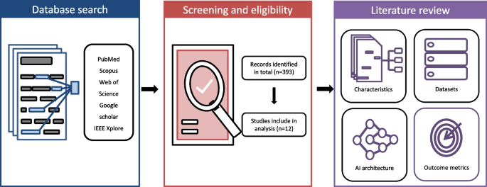
The workflow of this study is shown in Fig. 1. We followed the reporting recommendations specified in the Preferred Reporting Items for Systematic Reviews and Meta-Analyses for Scoping Reviews (PRISMA-ScR) guidelines [19] to convey the findings of this investigation. The specific review question was the applications and performance of AI in dental crown prostheses.
Our literature exploration was based on the PICO (problem/patient/population, intervention/indicator, comparison, and outcome) elements [20].
-
1.
Population: Images and other data types for dental crown prostheses used in prosthodontic rehabilitation.
-
2.
Intervention/Comparison: AI models for diagnosis, prognosis assessment, and treatment procedures compared to reference standards.
-
3.
Outcome: Any type of performance measurement.
Literature search
An electronic literature search was conducted in January 2024 across five databases: PubMed, Scopus, Web of Science, Google Scholar, and IEEE Xplore. We focused on literature published after 2010 to capture the most relevant and recent advancements in deep learning applications in dentistry. Manual searches and searches for gray literature were not conducted.
Search terms were developed and combined by two reviewers (H.J.K. and Y.L.K.) with reference to previous studies [16, 18]. Each category consists of a combination of Medical Subject Headings (MeSH) Terms and related dental terms with conjunctions: (“dental crown” OR “crown preparation” OR “fixed prosthesis” OR “prosthodontic” OR “dental prosthesis”) and (“artificial intelligence” OR “machine learning” OR “deep learning” OR “neural network”).
Eligibility criteria
The following inclusion criteria were employed in the selection of articles:
-
1.
Articles related to AI applications in dental crown prosthesis
-
2.
Articles composed in English and released between January 2010 and January 2024
Our exclusion criteria were:
-
1.
Articles that used AI for conditions not related to dental crown prosthesis
-
2.
Articles that did not report performance metrics such as accuracy
-
3.
Articles without the full text available
-
4.
Review articles and letters to the editor
Two reviewers (H.J.K. and Y.L.K.) assessed the titles and abstracts of articles based on pre-established inclusion and exclusion criteria. When titles and abstracts were deemed insufficient in providing necessary information, a thorough analysis of the entire text was conducted. Both reviewers diligently examined articles that were potentially relevant and collaboratively selected papers for further analysis. Any discrepancies in opinions were resolved through discussion.
Results
Selection of sources
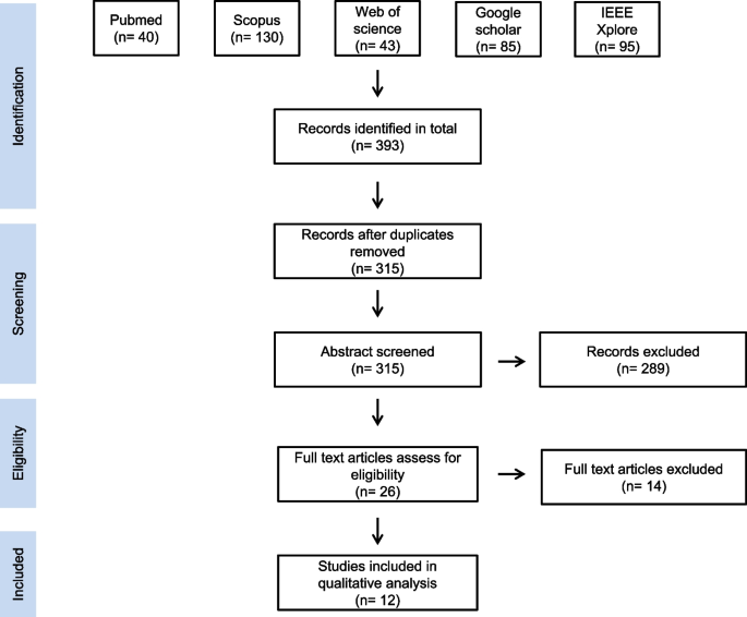
The flowchart in Fig. 2 outlines the article selection process adhering to PRISMA-ScR for this scoping review. Initially, a search of electronic literature produced 393 records, which was decreased to 315 after eliminating duplicate references. Following the examination of titles and abstracts, 26 studies underwent a more detailed review, after which 14 records were excluded for not meeting the inclusion criteria. Finally, 12 eligible publications were included in the qualitative review (Table 1). The evaluators unanimously agreed on the literature selection and categorization of publications.
Characteristics of the included studies
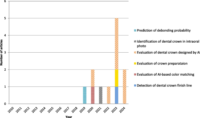
The AI-based applications included in this review were related to detection of dental crown finish line, evaluation of AI-based color matching, evaluation of crown preparation, evaluation of dental crown designed by AI, identification of dental crowns in intraoral photos, and prediction of debonding probability (Fig. 3). While the search was initially planned to encompass articles published between January 2010 and January 2024, the findings indicate a surge in the popularity of AI applications starting in 2019.
Datasets
The dataset size exhibited a wide range owing to the diversity in inputs and outcomes across study types (Table 2). Dataset size for included studies ranged from 12 to 8640. Studies using AI software included a small number of data (mean = 24.25). Since these studies do not learn new models, they do not require large datasets like existing deep learning studies. The most common types of data used were scanned digital images of the jaw and casts (n=10), and two studies used intraoral photographs.
AI architecture
Three types of AI architecture were used in this body of literature. AI software was used most often (5 times). CNN was used four times, and GAN was used three times. Among AI software, CNN and GAN algorithms were used in two studies. However, in the remaining three documents, the name of the software was provided, although the specific algorithm was not disclosed.
Excluding studies where the algorithm could not be confirmed, in designs using AI, the program used only 3D-GAN or used it in combination with CNN. In other types of studies, CNNs, which are widely used in image classification and object detection, were used. In two studies using a combination of CNN and GAN, CNN was used to extract the preparation tooth and set the margin line, and then GAN was used to create the outer surface.
Outcome metrics
With a scoping review, there is considerable heterogeneity in data forms and methodologies, leading to diverse outcome metrics. In studies evaluating AI design, RMS was used as an evaluation indicator in six of the studies. In these studies, mean deviation to evaluate positional accuracy and cusp angle to evaluate crown shape were used as evaluation indicators. Working time was also included in two studies to compare with existing programs or traditional methods. When an object detection algorithm was used, Intersection over union (IoU) was used as an evaluation index. Accuracy, precision, recall, and F-score were used in one prediction study.
Discussion
According to this scoping review, an increasing number of studies have employed AI for various tasks related to dental crown prostheses. To align with the rapid evolution of digital technologies, the investigation focused on the most recent 14 years. The application of AI in the fabrication and evaluation of dental crowns can contribute to increased efficiency for prosthodontists, raising expectations for improved productivity. The application of AI to the following fields related to dental crown prosthesis was reviewed.
Dental crown prosthesis designed by AI
The production of crown prostheses has been separated into traditional wax-up methods and digital approaches using CAD/CAM systems. Recently, there has been a widespread adoption of CAD/CAM methodologies, particularly in conjunction with the popularization of zirconia usage. Notably, these methods report high levels of accuracy and success rates [33]. In the CAD/CAM process, there have been efforts to enhance the speed and precision of prosthesis design through the application of AI [34].
Seven studies evaluated the feasibility of AI models to design dental crown prostheses [21,22,25,28,29,31,32. Four studies used AI software [25, 28, 31, 32], while three studies [21, 22, 29] employed 3D-GAN algorithms for model training. In six studies, integration of AI into the fabrication process demonstrated higher accuracy compared with traditional CAD or manually designed methods. One study [28] reported that knowledge-based AI, compared to human-designed CAD software, exhibited a higher occlusal profile discrepancy.
Two studies [25, 31] reported high time efficiency of AI-based programs. While traditional CAD work does not involve adding and modifying wax, it requires human thinking. Both wax-up and CAD processes are heavily influenced by the accumulated experience of dental technicians [35, 36]. However, AI-based operations minimize human intervention using automated calculations. Therefore, designing with AI significantly reduces the time spent on the design process, especially in cases with extensive restoration requirements.
Among the seven studies, four used commercially produced AI software, with two of them lacking specific mention of the program's algorithm [25, 28]. The absence of detailed information on the algorithms and metrics used for training the models in these studies acts as a limitation, emphasizing the need for transparency in the application of AI in dentistry, especially with commercially available software. Additionally, studies employing software had datasets ranging from 12 to 30 cases, posing a limitation in detecting statistical significance due to the potentially insufficient sample size. Despite these limitations, AI for dental crown design has the potential to significantly increase production efficiency by saving time. Considering the performance demonstrated in aspects such as morphology, internal fit, and occlusion, there is a promising outlook for the future utilization of AI in dentistry.
Detection of dental crown finish line
Choi et al. [23] compared the accuracy of hybrid software combined with deep learning with existing traditional CAD software in detecting the crown finish line. An accurate marginal fit is crucial for preventing microgaps. This, in turn, lowers the risk of caries and ensures that the restoration retains its function [37]. Recently, finish lines have been extracted and processed manually using CAD programs, but this is a repetitive and time-consuming process [38]. In Choi et al., as a result of evaluation using Hausdorff distance and chamfer distance, the hybrid system showed statistically more accurate results. This implies that a hybrid approach, integrating both deep learning and computer-aided design methods, may allow robust and precise extraction of finish lines with minimal adjustments required.
Evaluation of crown preparation
One of the most fundamental aspects in dental prosthodontics education is understanding and practicing the principles of tooth preparation [39]. However, evaluating students' tooth preparation outcomes in dental education can lack consistency due to factors such as subjective grading scales and insufficient inter-rater agreement. This difficulty hinders the provision of ongoing and reliable feedback [40, 41].
Han et al. [27] assessed the viability of software-based automated evaluation (SAE) with AI to evaluate abutment tooth preparation for single crowns. This was done through a comparison with a human-based digitally-assisted evaluation (DAE), which showed perfect intra-rater agreement and almost perfect inter-rater agreement with SAE. The findings of this study substantiate the credibility of SAE within prosthodontics education and propose its potential clinical utility for evaluating tooth preparation
Evaluation of AI-based color matching
A crucial aspect of the dental technician's role is replicating the natural color of teeth in dental prostheses. An experienced dental technician possesses the ability to precisely assess the authentic color. However, this proves to be a challenging task for a less-experienced dental technician [42].
Ueki et al. [26] extracted 62 images of patient teeth, which were annotated by experienced dental technicians. They then used a neural network to estimate the true color. The accuracy of the first candidate's output was six of 22 (27%), considerably lower than the desired level. However, the outputs for the second and third candidates encompassed 12 (55%) and 15 (68%) of the total 22 images, respectively. This affirmed accurate classification of certain colors.
One notable limitation of this study is the relatively small size of the image dataset used. To more accurately assess the potential of AI in shade selection, a substantial amount of training data is required.
Identification of dental crown in intraoral photo
In clinical situations, it is crucial for dentists to gather intraoral information about patients—a process that demands time and effort. Additionally, the effectiveness of this procedure relies on the dentist's knowledge and experience. Consequently, there is a demand for an automated system that can rapidly assess the intraoral situation.
Takahashi et al. [24] used a deep learning object detection method to recognize dental prostheses and restorations. In their study, ‘You Only Look Once version 3’ (YOLOv3) was used for object detection because it has shown high performance in other dental deep learning studies [43,44,45].
A satisfactory level of performance is typically associated with an IoU exceeding 0.7 [46, 47]. In the present investigation, the IoU was 0.76. Consequently, the proficiency of this learning system was high. In assessing the accuracy of the object detection model, the mAP is employed, with values above 0.7 considered favorable in previous research [48]. The mAP achieved in the present study was 0.80, supporting the learning system's commendable performance from a mAP perspective.
Irrespective of the overall count of objects across all images, there was a tendency for higher average precision (AP) scores in cases of metallic-colored prostheses, while tooth-colored prostheses exhibited a tendency toward lower AP scores. These findings suggest that the identification was influenced by the color distinctions between the natural teeth and prostheses.
Prediction of debonding probability
CAD/CAM composite resin (CR) crowns cemented to dentin often exhibit a propensity for debonding within one year, and the reported debonding rate for CAD/CAM CR crowns cemented on implant abutments stands at 80% within one year [49]. Inadequate preparation has been identified to contribute to debonding [50, 51].
Yamaguchi et al. [30] aimed to predict the debonding probability of CAD/CAM CR crowns using scanned images of prepared models employing convolutional neural networks. The reported prediction accuracy was 98.5%. Despite the good performance, this study acknowledges a limitation in explaining the primary factor contributing to debonding, stating that it was difficult to pinpoint the main cause.
Limitations
This study has several limitations. First, as a scoping review, it provides an overview of the current application of AI in dental crown prosthetics and assesses the need for further research, but it does not establish the reliability of existing knowledge as a systematic review would. Second, the reviewed literature was classified into six categories, but half of the articles are included in the category of "evaluation of dental crowns designed by AI," with only one article in each of the other categories. Lastly, some of the articles included in the review lack transparency as the algorithms used are not clearly specified.
In addition to the articles reviewed in this study, there are an increasing number of studies integrating next-generation technologies, such as robotics, into dental crown prosthesis [52, 53]. The advancement of these technologies, including AI, will bring continuous progress to the entire field of prosthodontics.
Conclusions
The number of studies applying AI to dental crown prostheses is gradually increasing. According to the results of this review, AI has the potential to increase efficiency in processes such as fabricating and evaluating dental crowns, with a high level of accuracy reported in most of the included studies. However, a significant number of studies focused on designing crowns using AI-based software, and these studies have limitations in that the study size was small and the algorithms used were sometimes not disclosed. The small number of AI-related studies means that AI application to dental crowns is needed in more diverse aspects. Additionally, research involving a large number of patients and data in various clinical situations is needed. AI study, by its nature, uses a variety of methods and evaluation metrics. Therefore, standardized protocols for reporting and evaluating AI studies are needed to increase the evidence and effectiveness.
Availability of data and materials
All data generated or analysed during this study are included in this published article.
Abbreviations
- AI:
-
Artificial intelligence
- CAD:
-
Computer-aided design
- CAD/CAM:
-
Computer-aided design/computer-aided manufacturing
- CNN:
-
Convolutional neural network
- CR:
-
Crown composite resin crown
- DAE:
-
Digitally-assisted evaluation
- DCGAN:
-
Deep convolutional generative adversarial network
- DCPR-GAN:
-
Two-stage deep generative adversarial network
- GAN:
-
Generative adversarial network
- SAE:
-
Software-based automated evaluation
- IoU:
-
Intersection over union
- RMS:
-
Root mean square error
References
Briganti G, Le Moine O. Artificial Intelligence in Medicine: Today and Tomorrow. Front Med (Lausanne). 2020;7:27.
Topol EJ. High-performance medicine: the convergence of human and artificial intelligence. Nat Med. 2019;25:44–56.
Paul D, Sanap G, Shenoy S, Kalyane D, Kalia K, Tekade RK. Artificial intelligence in drug discovery and development. Drug Discov Today. 2021;26:80–93.
Schwendicke F, Samek W, Krois J. Artificial intelligence in dentistry: chances and challenges. J Dent Res. 2020;99:769–74.
Ahmed N, Abbasi MS, Zuberi F, Qamar W, Halim MSB, Maqsood A, et al. Artificial intelligence techniques: analysis, application, and outcome in dentistry-a systematic review. Biomed Res Int. 2021;9751564. https://doi.org/10.1155/2021/9751564.
Shan T, Tay FR, Gu L. Application of artificial intelligence in dentistry. J Dent Res. 2021;100:232–44.
Lee JH, Kim DH, Jeong SN, Choi SH. Detection and diagnosis of dental caries using a deep learning-based convolutional neural network algorithm. J Dent. 2018;77:106–11.
Zhang X, Liang Y, Li W, Liu C, Gu D, Sun W, et al. Development and evaluation of deep learning for screening dental caries from oral photographs. Oral Dis. 2022;28:173–81.
Aminoshariae A, Kulild J, Nagendrababu V. Artificial intelligence in endodontics: current applications and future directions. J Endod. 2021;47:1352–7.
Ahmed ZH, Almuharib AM, Abdulkarim AA, Alhassoon AH, Alanazi AF, Alhaqbani MA, et al. Artificial intelligence and its application in endodontics: a review. J Contemp Dent Pract. 2023;24:912–7.
Nishimoto S, Sotsuka Y, Kawai K, Ishise H, Kakibuchi M. Personal computer-based cephalometric landmark detection with deep learning, using cephalograms on the internet. J Craniofac Surg. 2019;30:91–5.
Sukegawa S, Yoshii K, Hara T, Yamashita K, Nakano K, Yamamoto N, et al. Deep neural networks for dental implant system classification. Biomolecules. 2020;10(7):984.
Kong HJ, Eom SH, Yoo JY, Lee JH. Identification of 130 dental implant types using ensemble deep learning. Int J Oral Maxillofac Implants. 2023;38:150–6.
Alqutaibi AY, Algabri RS, Elawady D, Ibrahim WI. Advancements in artificial intelligence algorithms for dental implant identification: A systematic review with meta-analysis. J Prosthet Dent. 2023;S0022–3913(23):00783–7.
Castiglioni I, Rundo L, Codari M, Di Leo G, Salvatore C, Interlenghi M, et al. AI applications to medical images: from machine learning to deep learning. Phys Med. 2021;83:9–24.
Arsiwala-Scheppach LT, Chaurasia A, Müller A, Krois J, Schwendicke F. Machine learning in dentistry: a scoping review. J Clin Med. 2023;12:937.
Blatz MB, Chiche G, Bahat O, Roblee R, Coachman C, Heymann HO. Evolution of aesthetic dentistry. J Dent Res. 2019;98:1294–304.
Revilla-León M, Gómez-Polo M, Vyas S, Barmak AB, Gallucci GO, Att W, et al. Artificial intelligence models for tooth-supported fixed and removable prosthodontics: a systematic review. J Prosthet Dent. 2023;129:276–92.
Tricco AC, Lillie E, Zarin W, et al. PRISMA extension for scoping reviews (PRISMA-ScR): checklist and explanation. Ann Intern Med. 2018;169:467–73.
Eriksen MB, Frandsen TF. The impact of patient, intervention, comparison, outcome (PICO) as a search strategy tool on literature search quality: a systematic review. J Med Libr Assoc. 2018;106:420–31.
Chau RCW, Hsung RT, McGrath C, Pow EHN, Lam WYH. Accuracy of artificial intelligence-designed single-molar dental prostheses: a feasibility study. J Prosthet Dent. 2023;S0022–3913(22):00764–8.
Tian S, Wang M, Dai N, Ma H, Li L, Fiorenza L, et al. Dcpr-gan: dental crown prosthesis restoration using two-stage generative adversarial networks. IEEE J Biomed Health Inform. 2022;26:151–60.
Choi J, Ahn J, Park JM. Deep learning-based automated detection of the dental crown finish line: an accuracy study. J Prosthet Dent. 2023;S0022–3913(23):00769–2.
Takahashi T, Nozaki K, Gonda T, Mameno T, Ikebe K. Deep learning-based detection of dental prostheses and restorations. Sci Rep. 2021;11:1960.
Liu CM, Lin WC, Lee SY. Evaluation of the efficiency, trueness, and clinical application of novel artificial intelligence design for dental crown prostheses. Dent Mater. 2024;40:19–27.
Ueki K, Wakamatsu H, Hagiwara Y. Evaluation of dental prosthesis colors using a neural network. 2020 IEEE 5th International Conference on Signal and Image Processing, Nanjing, China. 2020;210–214. https://ieeexplore.ieee.org/document/9339381. Accessed 15 Feb 2024.
Han S, Yi Y, Revilla-León M, Yilmaz B, Yoon HI. Feasibility of software-based assessment for automated evaluation of tooth preparation for dental crown by using a computational geometric algorithm. Sci Rep. 2023;13:11847.
Chen Y, Lee JKY, Kwong G, Pow EHN, Tsoi JKH. Morphology and fracture behavior of lithium disilicate dental crowns designed by human and knowledge-based AI. J Mech Behav Biomed Mater. 2022;131:105256.
Ding H, Cui Z, Maghami E, Chen Y, Matinlinna JP, Pow EHN, et al. Morphology and mechanical performance of dental crown designed by 3D-DCGAN. Dent Mater. 2023;39:320–32.
Yamaguchi S, Lee C, Karaer O, Ban S, Mine A, Imazato S. Predicting the debonding of cad/cam composite resin crowns with ai. J Dent Res. 2019;98:1234–8.
Cho JH, Yi Y, Choi J, Ahn J, Yoon HI, Yilmaz B. Time efficiency, occlusal morphology, and internal fit of anatomic contour crowns designed by dental software powered by generative adversarial network: a comparative study. J Dent. 2023;138:104739.
Cho JH, Çakmak G, Yi Y, Yoon HI, Yilmaz B, Schimmel M. Tooth morphology, internal fit, occlusion and proximal contacts of dental crowns designed by deep learning-based dental software: a comparative study. J Dent. 2024;141:104830.
Leitão CIMB, Fernandes GVO, Azevedo LPP, Araújo FM, Donato H, Correia ARM. Clinical performance of monolithic CAD/CAM tooth-supported zirconia restorations: systematic review and meta-analysis. J Prosthodont Res. 2022;66:374–84.
Bernauer SA, Zitzmann NU, Joda T. The use and performance of artificial intelligence in prosthodontics: a systematic review. Sensors (Basel). 2021;21:6628.
Teng TY, Wu JH, Lee CY. Acceptance and experience of digital dental technology, burnout, job satisfaction, and turnover intention for Taiwanese dental technicians. BMC Oral Health. 2022;22:342.
Litzenburger AP, Hickel R, Richter MJ, Mehl AC, Probst FA. Fully automatic CAD design of the occlusal morphology of partial crowns compared to dental technicians’ design. Clin Oral Invest. 2013;17:491–6.
Larson TD. The clinical significance of marginal fit. Northwest Dent J. 2012;91:22.
Mai HN, Han JS, Kim HS, Park YS, Park JM, Lee DH. Reliability of automatic finish line detection for tooth preparation in dental computer aided software. J Prosthodont Res. 2023;67:138–43.
Liu L, Zhou R, Yuan S, Sun Z, Lu X, Li J, et al. Simulation training for ceramic crown preparation in the dental setting using a virtual educational system. Eur J Dent Educ. 2020;24:199–206.
Kateeb ET, Kamal MS, Kadamani AM, Abu Hantash RO, Abu Arqoub MM. Utilising an innovative digital software to grade pre-clinical crown preparation exercise. Eur J Dent Educ. 2017;21:220–7.
Feil PH, Gatti JJ. Validation of a motor skills performance theory with applications for dental education. J Dent Educ. 1993;57:628–33.
Hardan L, Bourgi R, Cuevas-Suárez CE, Lukomska-Szymanska M, Monjarás-Ávila AJ, Zarow M, et al. Novel trends in dental color match using different shade selection methods: a systematic review and meta-analysis. Materials (Basel). 2022;15:468.
Kong HJ, Yoo JY, Lee JH, Eom SH, Kim JH. Performance evaluation of deep learning models for the classification and identification of dental implants. J Prosthet Dent. 2023;S0022–3913(23):00467–5.
Almalki YE, Din AI, Ramzan M, Irfan M, Aamir KM, Almalki A, et al. Deep learning models for classification of dental diseases using orthopantomography x-ray opg images. Sensors (Basel). 2022;22:7370.
Yilmaz S, Tasyurek M, Amuk M, Celik M, Canger EM. Developing deep learning methods for classification of teeth in dental panoramic radiography. Oral Surg Oral Med Oral Pathol Oral Radiol. 2023;S2212–4403(23):00116–5.
Tao R, Gavves E and Smeulders AWM. Siamese instance search for tracking. The IEEE Conference on Computer Vision and Pattern Recognition. 2016:1420–9. https://www.computer.org/csdl/proceedings-article/cvpr/2016/8851b420/12OmNscOUb0. Accessed 15 Feb 2024.
Behpour S, Kitani KM and Ziebart BD. Adversarially optimizing intersection over union for object localization tasks. CoRR. arXiv :1710.07735 2017. https://www.researchgate.net/publication/320582581_Adversarially_Optimizing_Intersection_over_Union_for_Object_Localization_Tasks. Accessed 15 Feb 2024.
Zhao ZQ, Zheng P, Xu ST, Wu X. Object detection with deep learning: a review. IEEE Trans Neural Netw Learn Syst. 2019. https://doi.org/10.48550/arXiv.1807.05511.
Schepke U, Meijer HJ, Vermeulen KM, Raghoebar GM, Cune MS. Clinical bonding of resin nano ceramic restorations to zirconia abutments: a case series within a randomized clinical trial. Clin Implant Dent Relat Res. 2016;18:984–92.
Rosentritt M, Preis V, Behr M, Krifka S. In-vitro performance of cad/cam crowns with insufficient preparation design. J Mech Behav Biomed Mater. 2019;90:269–74.
Yang Y, Yang Z, Zhou J, Chen L, Tan J. Effect of tooth preparation design on marginal adaptation of composite resin CAD-CAM onlays. J Prosthet Dent. 2020;124:88–93.
Alqutaibi AY, Hamadallah HH, Alturki KN, Aljuhani FM, Aloufi AM, Alghauli MA. Practical applications of robots in prosthodontics for tooth preparation and denture tooth arrangement: a scoping review. J Prosthet Dent. 2024;S0022–3913(24):00120–3.
Alqutaibi AY, Hamadallah HH, Abu Zaid B, Aloufi AM, Tarawah RA. Applications of robots in implant dentistry: a scoping review. J Prosthet Dent. 2023;S0022–3913(23):00770–9.
Acknowledgements
Not applicable.
Funding
This study was supported by Wonkwang University in 2024.
Author information
Authors and Affiliations
Contributions
HJK conceived the study. All authors developed the review protocol. HJK and YLK did the literature search and assessed the quality of included studies. All authors read, commented critically and approved the final manuscript.
Corresponding author
Ethics declarations
Ethics approval and consent to participate
Not applicable.
Consent for publication
Not applicable.
Competing interests
The authors declare no competing interests.
Additional information
Publisher’s Note
Springer Nature remains neutral with regard to jurisdictional claims in published maps and institutional affiliations.
Rights and permissions
Open Access This article is licensed under a Creative Commons Attribution-NonCommercial-NoDerivatives 4.0 International License, which permits any non-commercial use, sharing, distribution and reproduction in any medium or format, as long as you give appropriate credit to the original author(s) and the source, provide a link to the Creative Commons licence, and indicate if you modified the licensed material. You do not have permission under this licence to share adapted material derived from this article or parts of it. The images or other third party material in this article are included in the article’s Creative Commons licence, unless indicated otherwise in a credit line to the material. If material is not included in the article’s Creative Commons licence and your intended use is not permitted by statutory regulation or exceeds the permitted use, you will need to obtain permission directly from the copyright holder. To view a copy of this licence, visit http://creativecommons.org/licenses/by-nc-nd/4.0/.
About this article
Cite this article
Kong, HJ., Kim, YL. Application of artificial intelligence in dental crown prosthesis: a scoping review. BMC Oral Health 24, 937 (2024). https://doi.org/10.1186/s12903-024-04657-0
Received:
Accepted:
Published:
DOI: https://doi.org/10.1186/s12903-024-04657-0


